Injection Site Sarcoma in Cats: Causes, Treatment & Prognosis
December 12, 2016
What is a Feline Injection Site Sarcoma?
Injection site sarcomas are tumors of connective tissue origin in the skin and subcutaneous region in cats. Subtypes include osteosarcoma (bone origin), fibrosarcoma (fibrous tissue origin), chondrosarcoma (cartilage origin), etc.; however, these tumors types tend to behave similarly and are therefore treated in the same manner. These tumors can occur anywhere on the body, but are often located in areas where injections are frequently administered including: between the shoulder blades, on the legs, and in the hip regions.
What causes Feline Injection Site Sarcomas?
In the 1990s, several epidemiologic studies demonstrated a correlation with the development of these tumors and prior administration of injections (especially vaccines) under the skin at that site. Vaccines that have been most implicated as a cause include the killed rabies vaccine and feline leukemia virus (FeLV) vaccine, however, other vaccines and injections have been associated with tumor development. The time for these tumors to occur ranges from 4 weeks to as long as 10 years after injection. In 1996, the Vaccine-Associated Feline Sarcoma Task Force (VAFSTF) was formed and created the vaccination methods we use today. The need to prevent infectious diseases is now balanced with the risk of these injection-site associated sarcomas, and vaccine recommendations are made for each pet on an individual basis. The true incidence of injection-associated sarcomas in the cat is unknown but is believed to be between 1/10,000-10/10,000 cats, a much higher incidence rate than those seen in cats with non-vaccine related fibrosarcomas.
How is it Diagnosed?
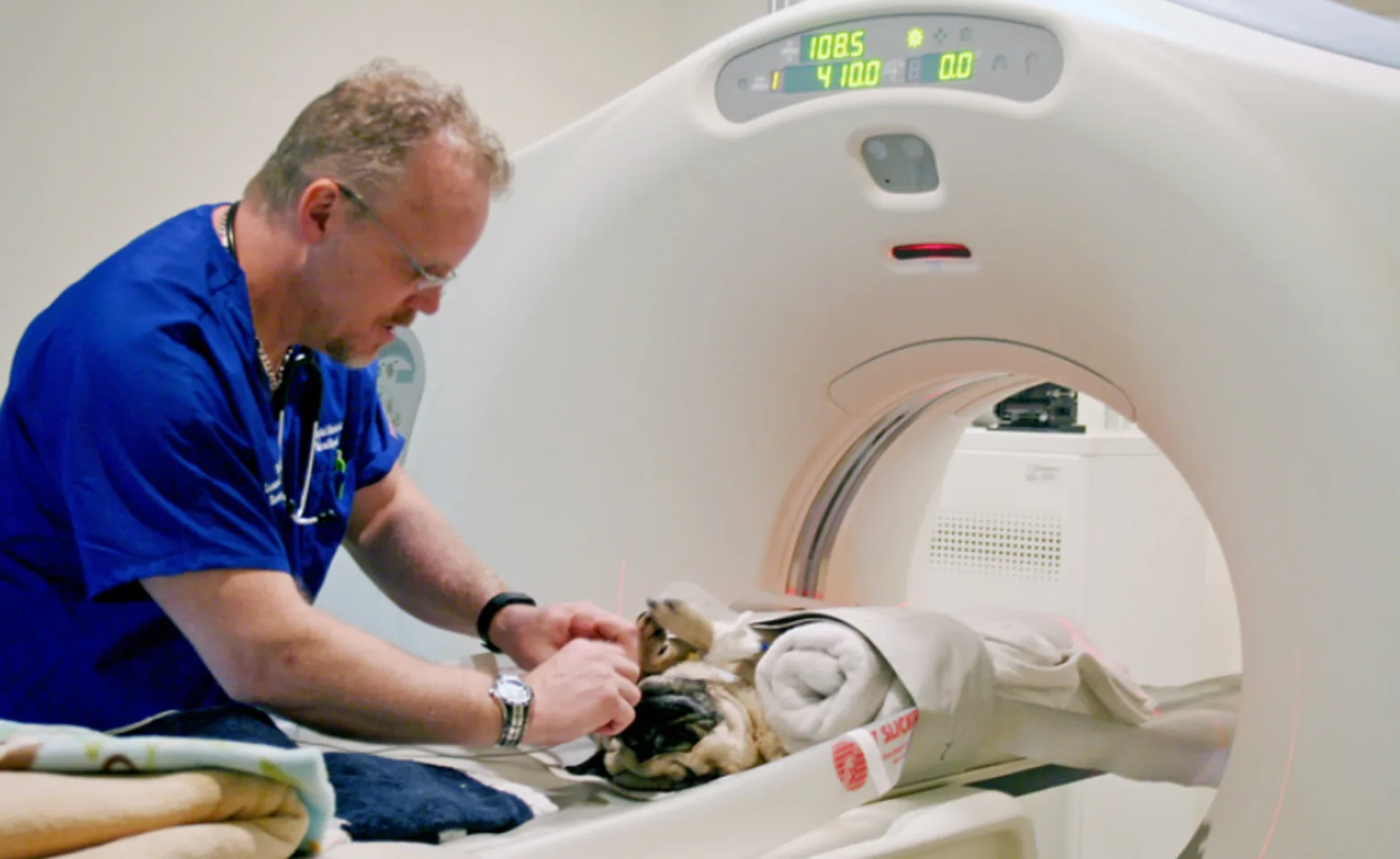
The “3-2-1” rule was created by the VAFSTF and states that if a mass has been present for 3 months or more after vaccination, is greater than 2 cm in diameter, or increases in size 1 month after vaccination, it should be biopsied. Biopsies are needed for a definitive diagnosis, and it is recommended that a small incisional biopsy be performed first prior to surgical excision. Once a diagnosis has been made, a CT scan or an MRI is highly recommended to better evaluate the extent of the disease. With the results of the MRI or CT scan, the surgeon or radiation therapist can better plan their approach and treatment of the tumor for the best possible outcome.
Prior to surgery, a CBC, chemistry panel, and urinalysis are recommended to assess your pet’s overall health status. Although the overall metastatic potential for these tumors is low (between 10-35%), thoracic radiographs and regional lymph node assessment are recommended. Sarcomas typically metastasize to the lungs but can also spread to other sites including the skin, regional lymph nodes, liver, and pelvic region.
What are the Treatment Options?
- Surgery: This is the best chance for a cure if it is pre-operatively determined (with MRI or CT scan) that the mass can be successfully excised. Lesions located on the limbs have a better prognosis because they are more amenable to surgery. The first surgery is the most important, and it is possible to obtain a cure if the surgery completely removes the tumor and all its microscopic tendrils (i.e. the surgical margins are clean of cancerous cells). This often requires at least 4-5 cm margins taken surgically in all directions around the initial tumor and is best pursued by a well-versed surgeon. Multiple surgeries have a poorer overall prognosis and are associated with a 50% higher risk of local disease recurrence and regional/distant metastasis. If the surgical margins contain cancerous cells (unclean surgical margins), or if surgical margins are narrow (2cm or less) the chance of recurrence is high. Additional adjuvant therapy is therefore recommended.
- Radiation Therapy: This can be used either preoperatively or postoperatively. Radiation therapy combined with surgery helps to improve tumor control, but prognosis is dependent on whether microscopic or macroscopic disease exists. The goal of pre-operative radiation therapy is to decrease the size (sterilize the cells at the edges) of the tumor so that complete surgical removal is possible. A smaller radiation field and lower total radiation dose is required when radiation is used in the pre-operative setting. Post-operative radiation therapy is used when complete surgical excision is not achieved (unclean or narrow surgical margins). Radiation therapy may also be an option when surgical resection is not possible.
- Chemotherapy: Chemotherapy’s role has not yet been determined, but early studies are promising. We may recommend chemotherapy pre-operatively in the hopes of decreasing the size of the tumor so that complete surgical resection is possible, or post-operatively for higher grade tumors. Chemotherapy is generally well tolerated by cats, with minimal side effects.
- Small Molecule Inhibitor: There is anecdotal evidence that a class of drugs called receptor tyrosine kinase inhibitors that have activity against the cellular receptor for PDGF may help stabilize and or decrease the size of an unresectable tumor. It has not been evaluated with any rigor for the treatment of feline sarcomas, however, the mechanisms of action of this compound including cell signal inhibition (primarily PDGF-R inhibition), inhibiting tumor cell division, promoting tumor cell death, and decreased tumor mediated angiogenesis is the rationale for its consideration. Side effects associated with this therapy are not known in cats but may include gastrointestinal disturbances, decreased appetite, white blood cell suppression, lethargy, and anemia.
What is the Prognosis?
Unfortunately, this tumor is very difficult to control because of the small tentacles that project out from the main mass. These tentacles are the most common cause of recurrence. Unfortunately, recurrence and/or the presence of a large unresectable mass are the most common cause of death in these cats. Fortunately, there are several treatment options that are available at this time, and they can be used in combination to give your pet the best chance for long-term tumor control. One of the most important features of this tumor is early detection, because when caught early enough, surgery or surgery combined with radiation, can be curative.